This Understanding ARIA portal provides medical resources for neurologists, radiologists, and other healthcare professionals on amyloid-related imaging abnormalities (ARIA) in multiple learning formats including modules, expert video lectures, infographics, key publication summaries, and links to other educational materials.


Understanding ARIA
ARIA presents as edema/effusion (ARIA-E) and hemosiderin deposition due to microhemorrhage, superficial siderosis, or intracerebral hemorrhage (ARIA-H). ARIA-H, and less commonly ARIA-E, can occur spontaneously in patients with Alzheimer’s disease. It is thought to be related to underlying cerebral amyloid angiopathy (CAA). The risk of ARIA is increased with the use of monoclonal antibodies that remove amyloid plaques in patients with Alzheimer’s disease.
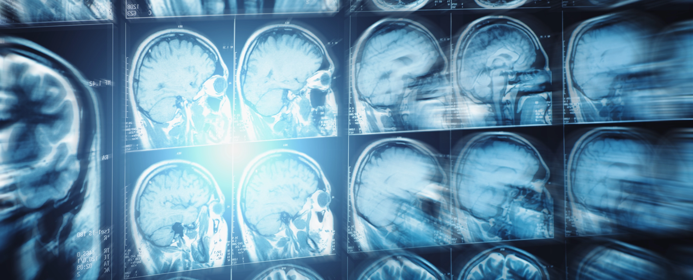
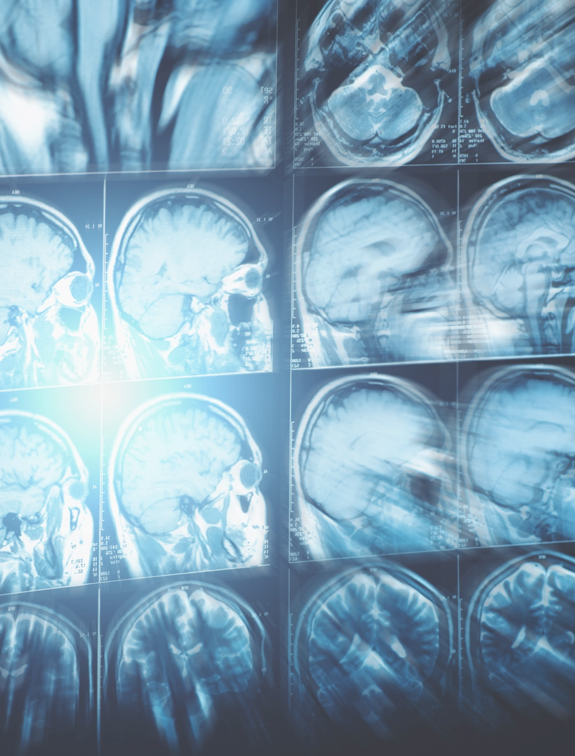
Magnetic resonance imaging (MRI) is used to monitor and diagnose ARIA. The occurrence of characteristic amyloid-related MRI signal changes requires careful consideration by the treating neurologist and radiologist. ARIA is often asymptomatic and usually resolves without intervention or treatment modification. When intervention or treatment modification is required, ARIA and any associated symptoms usually resolve, but serious events can occur.
